Microtia

What Is Microtia?
The outer ear is made up of cartilage, a connective tissue that develops in the womb, and in some cases, the growth of this tissue fails to progress properly. Microtia is considered a rare condition; however, the exact incidence and prevalence of microtia worldwide is unknown. It's estimated that 1 in 5,000 people is born with this condition.

General Information About Microtia
General Information
About Microtia
Microtia is associated with varying degrees of deformity:
- Grade I: Mild deformity
- Grade II: Small Ear
- Grade III: Sausage or Peanut shaped ear (most common)
- Grade IV: Anotia (absent ear).
Microtia statistics:
- Grade I: Mild deformity
- Grade II: Small Ear
- Grade III: Sausage or Peanut shaped ear (most common)
- Grade IV: Anotia (absent ear).
The Procedure: Rib Cartilage Framework for Microtia Reconstruction
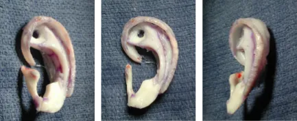
The result you can achieve through ear reconstruction depends on your surgeon’s artistic ability to sculpt the new ear from cartilage. The process begins with careful planning using measurements, patterns, molds, and plaster casts made from the ear on the opposite side.
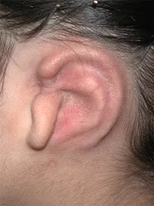
Stage I
During the first surgery, the patient’s own rib cartilage is carefully carved to make a framework that is implanted beneath the skin.
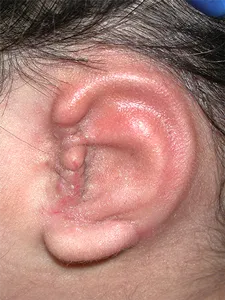
Stage II
Usually the earlobe is rotated into the proper position during the second stage of the reconstruction process.
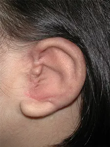
Stage III
During the third stage, the ear is elevated and a skin graft is placed behind the cartilage to create a space behind the ear.
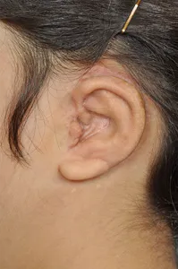
Stage IV
The forth stage typically includes refining the central cup of the ear (concha) and the protective door over the ear canal (tragus). A multiple stage approach is used to provide anatomic results.
Patient Photo Gallery
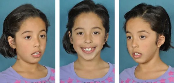
The photos to the left shows a seven-year-old girl, who had classic grade III (lobular) microtia, after four stages of ear reconstruction.
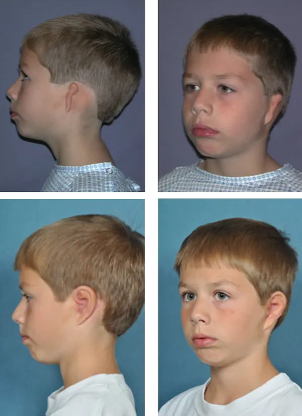
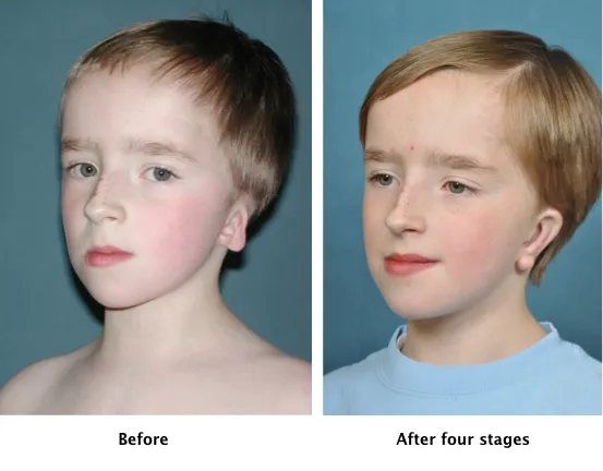
This eight-year-old boy has classic Grade III (Lobular) Microtia The top two pictures show him before his reconstructive surgery and the bottom two pictures show his results after four stages of microtia ear reconstruction.
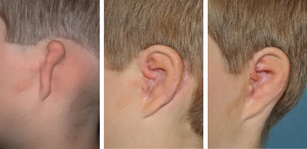
This eight-year-old boy had a classic Grade III microtia with atresia. The first picture on the left shows the boy’s ear before reconstructive surgery and the two pictures on the right show his results after the four stages of microtia repair.
This seven-year-old girl also had a Grade III (Lobular) microtia with atresia. The upper two photos show the patient prior to reconstructive ear surgery and the lower two photos demonstrate her results.
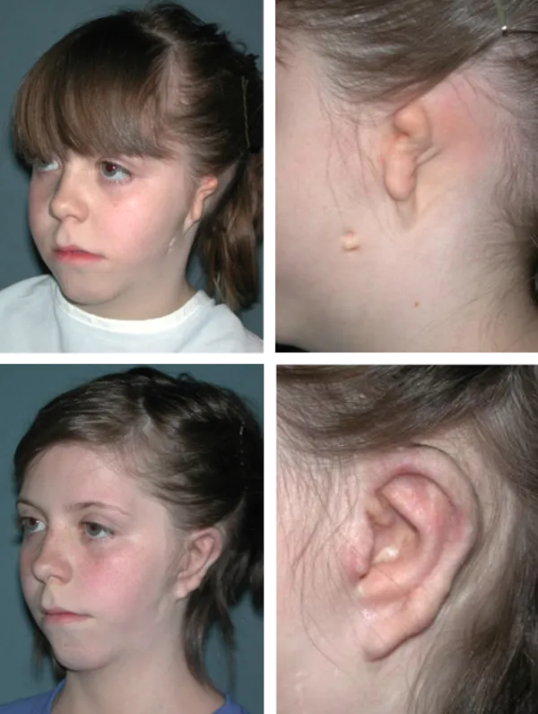

This seven-year-old girl also had a Grade III (Lobular) microtia with atresia. The upper two photos show the patient prior to reconstructive ear surgery and the lower two photos demonstrate her results.
Timing
Microtia reconstruction surgery can begin as early as age six. At this age the ear is approximately 85% of its adult size and there is enough rib cartilage to sculpt the new ear. This is also the time when the child is becoming aware of the deformity, often due to teasing at school. At this age, the child is more capable of caring for the ear after the operation.
Older patients can start the reconstruction process immediately following the loss of an ear due to cancer or as the result of an accident. In all cases, carving a new framework from the patient’s own rib cartilage will provide the best chance of success.
Secondary Ear Reconstruction
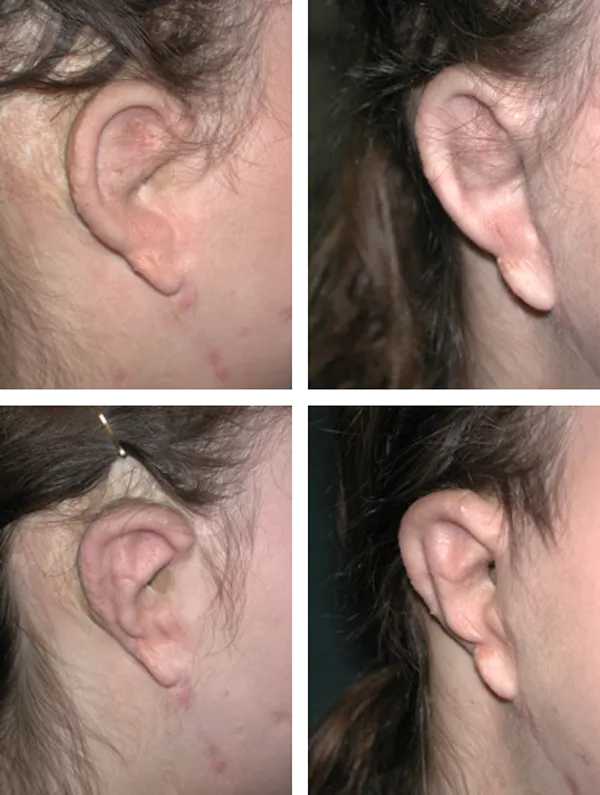
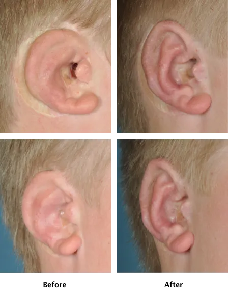
In these pictures, the top row shows the patient before secondary ear reconstruction. The patient had two Medpor frameworks placed in her ear by another surgeon and had developed an infection in the synthetic framework, which was causing persistent drainage.
c
The lower two pictures show the patient after secondary ear reconstruction. In these pictures the Medpor framework has been replaced by rib cartilage from the patient’s own rib tissue. The patient has had no graft loss, infection, or drainage during the three years since surgery.
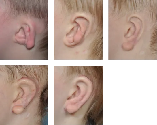
In these pictures, this seven-year-old boy had Dr. Grant A. Fairbanks perform a secondary ear reconstruction, since he wasn’t happy with the results he received with another surgeon. In the top-left photo, you can see how the ear looked before Dr. Fairbanks worked on the patient. The top-middle picture shows the new rib cartilage framework positioned under the skin. The top-right photo shows the earlobe after it had been moved forward (typically, the movement is done from front to back). The bottom-left photo shows the elevation of the ear from the scalp and the placement of a skin graft behind the ear. The last picture shows the final elevation using a cartilage graft placed behind the ear.
The picture on the left shows the results achieved by another surgeon. The patient was unhappy with these results, so he had Dr. Grant A. Fairbanks redo the procedure. The patient was pleased with the results of his secondary ear reconstruction, which are shown on the right.
This sixteen-year-old male had a procedure done by another surgeon. In the secondary ear reconstruction, Dr. Grant A. Fairbanks was able to achieve more detail in the ear and the patient has had no infection or graft loss and the ear is maintaining a good shape and size.
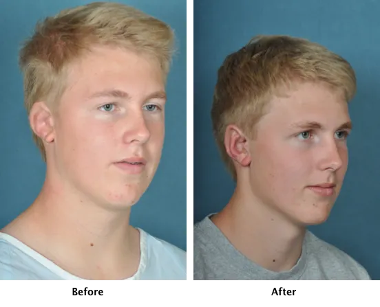
The picture on the left shows the results achieved by another surgeon. In the picture on the right, you can see the revised ear has more detail and a better shape and size.
The Procedure:
Rib Cartilage Framework
For Microtia Reconstruction

The result you can achieve through ear reconstruction depends on your surgeon’s artistic ability to sculpt the new ear from cartilage. The process begins with careful planning using measurements, patterns, molds, and plaster casts made from the ear on the opposite side.
Stage I
During the first surgery, the patient’s own rib cartilage is carefully carved to make a framework that is implanted beneath the skin.

Stage II
Usually the earlobe is rotated into the proper position during the second stage of the reconstruction process.

Stage III
During the third stage, the ear is elevated and a skin graft is placed behind the cartilage to create a space behind the ear.

Stage IV
The forth stage typically includes refining the central cup of the ear (concha) and the protective door over the ear canal (tragus). A multiple stage approach is used to provide anatomic results.

Contact
Grant A. Fairbanks

Disclaimer: This website is to be used for general information only and is not intended to provide medical advice. This site is not a substitute for consultations with Dr. Fairbanks.
Copyright 2024 Grant A. Fairbanks MD - Art of Plastic Surgery All Rights Reserved
